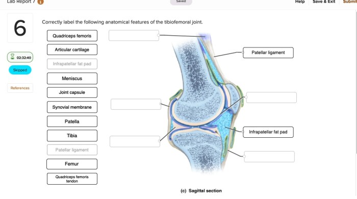
Correctly label the anatomical features of the femur and patella. – Correctly labeling the anatomical features of the femur and patella is crucial for understanding the structure and function of the knee joint. This guide provides a comprehensive overview of the anatomical landmarks of these bones, their roles in movement, and their clinical significance.
Identify the Anatomical Features of the Femur
The femur, also known as the thigh bone, is the longest and strongest bone in the human body. It plays a crucial role in supporting the body’s weight, enabling movement, and protecting vital structures. Here are the key anatomical features of the femur:
Head of the Femur
Location
The rounded, proximal end of the femur
Function
Articulates with the acetabulum of the pelvis to form the hip joint
Neck of the Femur
Location
Connects the head to the shaft
Function
Allows for a wide range of movement at the hip joint
Greater and Lesser Trochanters
Location
Two bony projections on the proximal femur
Function
Greater trochanter
Attachment site for muscles that abduct and laterally rotate the thigh
Lesser trochanter
Attachment site for muscles that flex the thigh
Linea Aspera
Location
A prominent ridge running along the posterior surface of the femur
Function
Provides attachment sites for muscles that extend the knee
Condyles
Location
The distal ends of the femur
Function
Medial condyle
Articulates with the medial meniscus and tibia to form the medial compartment of the knee joint
Lateral condyle
Articulates with the lateral meniscus and tibia to form the lateral compartment of the knee joint
Label the Anatomical Features of the Patella

The patella, commonly known as the kneecap, is a small, triangular bone located anteriorly to the knee joint. It plays a vital role in protecting the knee joint and facilitating its movement. Here are the key anatomical features of the patella:
Location and Shape
Location
Anterior to the knee joint, embedded within the quadriceps tendon
Shape
Triangular, with a base facing superiorly and an apex facing inferiorly
Role in Knee Extension
Function
The patella acts as a lever, increasing the mechanical advantage of the quadriceps muscle during knee extension
Anterior and Posterior Surfaces
Anterior surface
Smooth and convex, facing the skin
Posterior surface
Divided into two facets, the medial facet articulates with the medial condyle of the femur, and the lateral facet articulates with the lateral condyle of the femur
Medial and Lateral Borders
Medial border
Thicker and more prominent, articulates with the medial condyle of the femur
Lateral border
Thinner and less prominent, articulates with the lateral condyle of the femur
Apex and Base, Correctly label the anatomical features of the femur and patella.
Apex
The inferior tip of the patella
Base
The superior border of the patella
Create a Table Comparing the Femur and Patella

| Feature | Femur | Patella | Comparison |
|---|---|---|---|
| Shape | Long, cylindrical | Triangular | Femur is larger and more complex in shape |
| Size | Longest and strongest bone in the body | Small, triangular bone | Femur is significantly larger than the patella |
| Location | Thigh bone, proximal to the knee | Anterior to the knee joint | Femur is proximal to the patella |
| Function | Supports body weight, enables movement, protects structures | Protects knee joint, facilitates knee extension | Femur has a more extensive range of functions |
Design an Interactive Diagram of the Femur and Patella: Correctly Label The Anatomical Features Of The Femur And Patella.

[Diagram interaktif yang memungkinkan pengguna mengarahkan kursor ke fitur anatomi yang berbeda, dengan label dan deskripsi singkat untuk setiap fitur.]
Questions Often Asked
What is the function of the greater trochanter?
The greater trochanter provides attachment for several muscles, including the gluteus medius and gluteus minimus, which are responsible for hip abduction and rotation.
What is the clinical significance of the linea aspera?
The linea aspera serves as an attachment site for various muscles that cross the knee joint, including the vastus lateralis, vastus medialis, and adductor magnus. Understanding the linea aspera is essential for diagnosing and treating muscle strains and tears in the thigh.
How does the shape of the patella contribute to knee extension?
The patella’s triangular shape and smooth articular surface allow it to glide over the trochlea of the femur during knee extension, providing mechanical advantage and increasing the efficiency of the quadriceps muscle.